
When the His electrode records the earliest activation, an origin in the anteroseptal region of the right atrium is suggested.

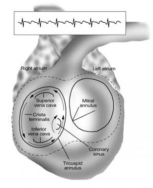
Intracardiac electrode activation sequences can indicate the site of origin of the AFL. Electrophysiologists depend on these electrograms as an initial step when performing procedures for any type of atrial flutter. This is particularly important in left atrial patterns, which can be observed in the electrograms of the distal coronary sinus. In all cases of atrial flutter, the activation patterns observed during flutter are essential for making an accurate diagnosis. More common atypical flutters include non-CTI-dependent right atrial flutter, peri-mitral flutter, roof-dependent left atrial flutter, and left atrial anterior wall flutter. Atypical flutters can occur in the right or left atrium. It is common for atypical flutter to transition to and from AF, and mapping studies have indicated that a range of circuits is feasible. ĮCG presentations of true atypical flutter are diverse. The most prevalent causes of non-CTI flutters are a history of ablation for atrial fibrillation (AF), prior cardiac surgery, particularly surgeries to correct congenital malformations or heart valve disease, and the presence of scar tissue within the atria that could have resulted from a prior atriotomy, atrial patch, or baffle. In these cases, the reentrant circuits follow pathways different than the cavotricuspid isthmus. Some types of atrial flutter are considered atypical, even though they involve a macroreentrant mechanism. Hence, it can be treated with ablation of this isthmus. Recognizing this arrhythmia is important similar to a typical counterclockwise flutter, the arrhythmia relies on the isthmus of tissue between the tricuspid valve annulus and the inferior vena cava. Clockwise CTI-dependent AFL can be detected on the surface ECG by observing P waves that are notched and upright in the inferior leads and inverted in lead V1. The cycle length is similar to that of a typical flutter, and intracardiac electrograms indicate that the reentrant circuit follows the same path but in the opposite direction. The most common variations among typical flutters seem to be clockwise CTI-dependent AFL. The wavefront also experiences a marked delay between its progression through the low right atrium and its arrival at the His location, during which it traverses the region of slow conduction in the isthmus. Moreover, CS activation occurs from the proximal to the distal sites, with the distal CS exhibiting a significantly delayed activation. Via catheter electrograms, the wavefront is initially detected by the proximal CS catheter, followed by the His catheter, and subsequently by the poles of the multipolar catheter located in the high, lateral, and low right atrium. On surface electrocardiogram (ECG) recordings, the P waves exhibit a sawtooth inverted pattern in the inferior leads, an inverted pattern in the high lateral leads, and an upright pattern in V1. A line of functional block can be observed along the crista terminals, precisely at the point where the ascending and descending signals collide. The electrical activity then flows back through the slow conduction zone between the tricuspid valve annulus and the CS. The signal is funneled into the narrow isthmus channel between the tricuspid valve annulus and the inferior vena cava, the cavotricuspid isthmus. There it travels between the tricuspid valve and the crista terminalis. The signal then crosses the roof of the right atrium and descends inferiorly and laterally. It then moves upwards towards the interatrial septum and depolarizes the posterior right atrium. The electrical activity propagates via a slow conduction zone between the tricuspid valve annulus and the coronary sinus (CS). This dysrhythmia is a macroreentrant counterclockwise circuit within the right atrium. Typical atrial flutter involves a circuit spanning the cavotricuspid isthmus (CTI).Ĭounterclockwise CTI-dependent Atrial FlutterĬounterclockwise CTI-dependent AFL is the most common atrial flutter variant. Atrial flutter may be classified as typical or atypical.


 0 kommentar(er)
